GLENDA: The ITEC Gynecologic Laparoscopy Endometriosis Dataset
GLENDA (Gynecologic Laparoscopy ENdometriosis DAtaset) comprises over 25 000 images taken from 400+ gynecologic laparoscopy surgeries and is purposefully created to be utilized for a variety of automatic content analysis problems in the context of Endometriosis recognition.
Endometriosis
Endometriosis is a benign but potentially painful condition among women in child bearing age involving the growth of uterine-like tissue in locations outside of the uterus. Corresponding lesions can be found in various positions and severities, often in multiple instances per patient requiring a physician to determine its extent. This most frequently is accomplished by calculating its magnitude via utilizing the combination of two popular classification systems, the revised American Society for Reproductive Medicine (rASRM) and the European Enzian scores. Endometriosis can not reliably identified by laymen, therefore, the dataset has been created with the help of medical experts in the field of endometriosis treatment.
Purposes
- binary/multi-label (endometriosis) classification
- detection/localization
- augmenting annotations: tracking over video segments
Overview
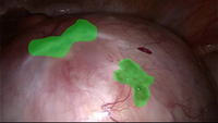
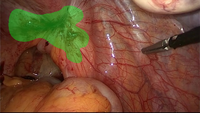
The dataset includes region-based annotations of 4 pathological endometriosis categories as well as non pathological counter example images. Annotations are created for single video frames that may be part of larger sequences comprising several consecutive frames (all showing the annotated condition). Frames can contain multiple annotations, potentially of different categories. Each single annotation is exported as a binary image (similar to below examples, albeit one image per annotation).
Categories
Example class frames with associated annotations, including original/annotated images with a representative sample video sequence for the category.
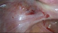
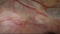
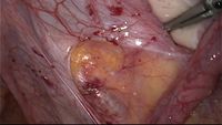
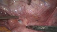
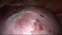
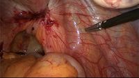
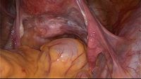
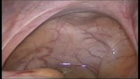
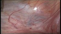
Peritoneum
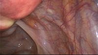
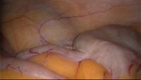
As one of the most frequently diagnosed types, peritoneal endometriosis is found on the peritoneum, i.e. the lining of the abdominal cavity, which occurs in a mixture of red, yellow and white colors.

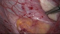

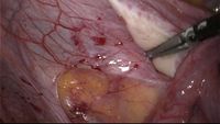

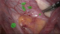

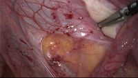
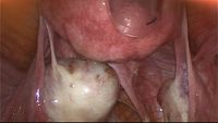

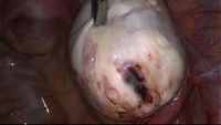
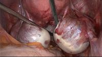
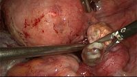
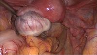
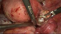
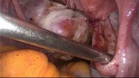
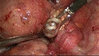
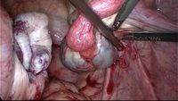
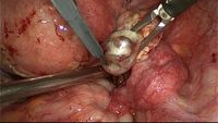
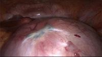
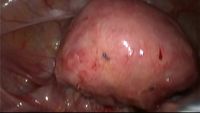
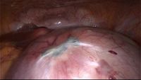
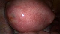
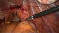
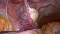
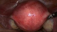
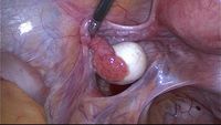
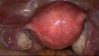
Ovary
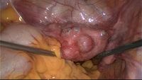
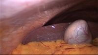
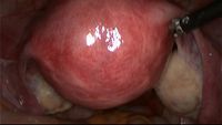
Endometriosis is as well very frequently found on ovaries, the outer capsule of which appears in a shade of white.


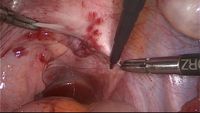
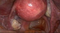
Uterus
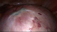
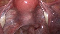
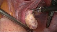
Uterine endometriosis or adenomyosis thickens the organ, which typically is colored in a red-tint.


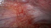
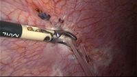
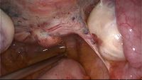
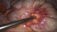
Deep infiltrating endometriosis (DIE)
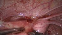
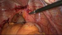
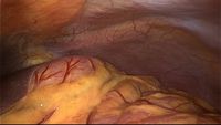
Non-shallow endometriosis that is found on specific locations such as the rectum, the rectovaginal space or uterine ligaments is described as Deep Infil- trating Endometriosis (DIE). Due to the variety of involved locations no distinct visual appearance can be attributed to this type of class, other than highlighting that typically the color spectrum in recorded laparascopic videos lacks green tones.

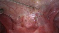


No pathology
Video sequences containing no visible pathology in relation to endometriosis are included in the dataset, providing counter examples to above categories. Since this class does not contain any region-based annotations, in addition to a sequence showing a non-pathological uterus, below listing particularly includes examples of several anatomical structures from above pathological classes (e.g. peritoneum and ovaries). Again it is not possible to make any assumptions about the color and shape of objects within this class, since it includes images covering most areas of the pelvic region.

Statistics
| Category* | annotations | annotated frames |
maximum annotations per frame |
sequences | total frames |
|---|---|---|---|---|---|
| peritoneum | 402 | 203 | 9 | 73 | 6470 |
| ovary | 51 | 48 | 2 | 15 | 2478 |
| uterus | 14 | 8 | 3 | 5 | 475 |
| DIE | 53 | 43 | 3 | 18 | 2821 |
| no pathology | 0 | 0 | 0 | 27 | 13 438 |
| Total | 520 | 302 | 9 (max. classes: 3) |
138 | 25 682 |
*Note: A sequence/keyframe/frame is attributed to a specific category if it is the dominant one in all of its corresponding annotations in terms of annotation count/area covered.
For further information about the dataset's organization see 'Readme.txt' within the download archive below.
Version History
All previous dataset revisions are listed here and their release pages are linked.
v1.5
- Removed all unannotated frames from dataset (for downloading the full version containing all frames in addition to the annotated keyframes, please visit the v1.0 page)
- Merged and color-coded all annotations of individual frames in the style of MS COCO format (annotations are now multi-colored pixel masks on a solid color background)
- Separated 'no pathology' image sequences from dataset containing annotated classes
v1.0 (this version)
- Initial release (with paper)
Disclaimer
The dataset is exclusively provided for scientific research purposes and as such cannot be used commercially or for any other purpose. If any other purpose is intended, you may directly contact the originator of the videos, Prof. Dr. Jörg Keckstein.
In addition, reference must be made to the following publication when this dataset is used in any academic and research reports:
A. Leibetseder, S. Kletz, K. Schoeffmann, S. Keckstein and J. Keckstein. 2020. GLENDA: Gynecologic Laparoscopy Endometriosis Dataset. In Proceedings of the 26th International Conference on Multimedia Modeling, MMM 2020. Springer, Cham.
@inproceedings{DBLP:conf/mmm/LeibetsederKSKK20,
    author    = {Andreas Leibetseder and
                 Sabrina Kletz and
                 Klaus Schoeffmann and
                 Simon Keckstein and
                 J{\"{o}}rg Keckstein},
    title     = {{GLENDA:} Gynecologic Laparoscopy Endometriosis Dataset},
    booktitle = {MultiMedia Modeling - 26th International Conference, {MMM} 2020, Daejeon,
                 South Korea, January 5-8, 2020, Proceedings, Part {II}},
    series    = {Lecture Notes in Computer Science},
    volume    = {11962},
    pages     = {439--450},
    publisher = {Springer},
    year      = {2020},
    url       = {https://doi.org/10.1007/978-3-030-37734-2\_36},
    doi       = {10.1007/978-3-030-37734-2\_36},
    timestamp = {Mon, 09 Nov 2020 15:46:42 +0100},
    biburl    = {https://dblp.org/rec/conf/mmm/LeibetsederKSKK20.bib},
    bibsource = {dblp computer science bibliography, https://dblp.org}
}
GLENDA is licensed under Creative Commons Attribution-NonCommercial 4.0 International (CC BY-NC 4.0,  ) and is created as well as maintained by Distributed Multimedia Systems Group of the Institute of Information Technology (ITEC) at Alpen-Adria Universität in Klagenfurt, Austria.
This license allows users of this dataset to copy, distribute and transmit the work under the following conditions:
- Attribution: You must give appropriate credit, provide a link to the license, and indicate if changes were made. You may do so in any reasonable manner, but not in any way that suggests the licensor endorses you or your use.
- Non-Commercial: You may not use the material for commercial purposes.
Download
If you agree to above conditions, you are free to download GLENDA (~1.7GB). Due to popular request a version without segments or non pathology images in an MS COCO format is available as well: GLENDA_MULTICOLOR (~21MB).